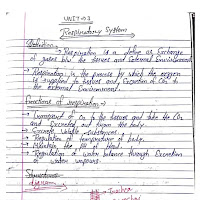
Download Respiratory System PDF, Notes, and PPT
Access detailed educational content on the Human Respiratory System. This free PDF download offers comprehensive notes and presentations covering the anatomy, physiology, and common pathologies of the respiratory system. Ideal for students of biology, medicine, and allied health sciences.
You can download the Respiratory System PDF or view the materials online. Our resources include clear explanations, diagrams of lung structures, mechanics of breathing, gas exchange, and more. Enhance your understanding with our free respiratory system notes and PPT slides!
- Keywords: Respiratory System PDF, anatomy notes, physiology PPT, download lung function PDF, breathing mechanics, gas exchange notes, free biology study material.
The Human Respiratory System: Anatomy, Physiology, and Function
The respiratory system is a complex network of organs and tissues primarily responsible for the process of breathing, or respiration. Its main function is to facilitate the exchange of gases between the body and the external environment, supplying oxygen to the blood and removing carbon dioxide. This system is vital for cellular respiration, which provides energy for all bodily functions.
Anatomy of the Respiratory System
The respiratory system can be divided into the upper respiratory tract and the lower respiratory tract, each with distinct structures and functions.
1. Upper Respiratory Tract
- Nose and Nasal Cavity: The primary entry point for air. Hairs and mucus in the nasal cavity filter, warm, and humidify inhaled air. The olfactory receptors for smell are also located here.
- Pharynx (Throat): A muscular funnel that serves as a common passageway for air (to the larynx) and food (to the esophagus). It is divided into three parts: nasopharynx, oropharynx, and laryngopharynx.
- Larynx (Voice Box): Connects the pharynx to the trachea. It contains the vocal cords, which produce sound when air passes over them. The epiglottis, a flap of cartilage, prevents food from entering the trachea during swallowing.
2. Lower Respiratory Tract
- Trachea (Windpipe): A cartilaginous tube that extends from the larynx into the chest, where it bifurcates. It is lined with ciliated epithelium and mucus-producing cells that trap and expel foreign particles.
- Bronchi: The trachea divides into two primary (main) bronchi, one for each lung. These further subdivide into secondary (lobar) bronchi, tertiary (segmental) bronchi, and progressively smaller bronchioles.
- Bronchioles: Smaller airways that branch off the bronchi. Terminal bronchioles mark the end of the conducting zone, leading to respiratory bronchioles, which are part of the respiratory zone.
- Alveoli: Tiny air sacs at the end of the respiratory bronchioles, arranged in clusters. They are the primary sites of gas exchange. The walls of alveoli are extremely thin and surrounded by a dense network of capillaries. Pulmonary surfactant, a substance produced by type II alveolar cells, reduces surface tension and prevents alveolar collapse.
- Lungs: The principal organs of respiration, located in the thoracic cavity on either side of the heart. The right lung typically has three lobes (superior, middle, inferior), while the left lung has two lobes (superior, inferior) to accommodate the heart. The lungs are enclosed by a double-layered membrane called the pleura (visceral pleura attached to the lung surface, parietal pleura lining the thoracic cavity), with a lubricating pleural fluid in between.
- Diaphragm and Intercostal Muscles: These are the primary muscles of respiration. The diaphragm is a large, dome-shaped muscle below the lungs, and the intercostal muscles are located between the ribs.
Physiology of Respiration
Respiration involves several key processes:
- Pulmonary Ventilation (Breathing): The mechanical process of moving air into and out of the lungs.
- Inspiration (Inhalation): An active process. The diaphragm contracts and flattens, and the external intercostal muscles contract, lifting the rib cage up and out. This increases the volume of the thoracic cavity, leading to a decrease in intrapulmonary pressure below atmospheric pressure, causing air to flow into the lungs.
- Expiration (Exhalation): Typically a passive process at rest. The diaphragm and external intercostal muscles relax, causing the thoracic cavity volume to decrease. This increases intrapulmonary pressure above atmospheric pressure, forcing air out of the lungs. Forced expiration involves contraction of internal intercostal and abdominal muscles.
- External Respiration (Gas Exchange in Lungs): The exchange of oxygen (O2) and carbon dioxide (CO2) between the air in the alveoli and the blood in the pulmonary capillaries. This occurs via simple diffusion across the respiratory membrane (alveolar and capillary walls). Oxygen moves from the alveoli (high O2 concentration) into the blood, and carbon dioxide moves from the blood (high CO2 concentration) into the alveoli.
- Transport of Respiratory Gases: Oxygen and carbon dioxide are transported in the blood.
- Oxygen Transport: Most oxygen (about 98.5%) binds to hemoglobin in red blood cells to form oxyhemoglobin. A small amount is dissolved in plasma.
- Carbon Dioxide Transport: Transported in three ways: dissolved in plasma (about 7-10%), bound to hemoglobin as carbaminohemoglobin (about 20%), and as bicarbonate ions (HCO3-) in the plasma (about 70%).
- Internal Respiration (Gas Exchange in Tissues): The exchange of O2 and CO2 between the systemic capillaries and tissue cells. Oxygen diffuses from the blood into the tissues, and carbon dioxide diffuses from the tissues into the blood.
Control of Breathing
Respiration is controlled by respiratory centers in the brainstem (medulla oblongata and pons). These centers regulate the rate and depth of breathing in response to various stimuli:
- Chemical Factors: Chemoreceptors (central in the medulla and peripheral in the carotid and aortic bodies) detect changes in blood levels of CO2, O2, and H+ (pH). Increased CO2 (hypercapnia) or H+ (acidosis), or significantly decreased O2 (hypoxia), stimulates an increase in ventilation. CO2 is the most potent chemical stimulus.
- Neural Factors: Higher brain centers (e.g., cerebral cortex) can voluntarily control breathing to some extent. Stretch receptors in the lungs (Hering-Breuer reflex) help prevent over-inflation.
Common Respiratory Conditions
The respiratory system is susceptible to various diseases and disorders, including:
- Infections: Common cold, influenza, bronchitis, pneumonia, tuberculosis.
- Obstructive Lung Diseases: Asthma, Chronic Obstructive Pulmonary Disease (COPD - encompassing chronic bronchitis and emphysema), cystic fibrosis. These are characterized by airflow limitation.
- Restrictive Lung Diseases: Pulmonary fibrosis, sarcoidosis. These are characterized by reduced lung expansion and total lung capacity.
- Lung Cancer: Malignant tumor growth in the lungs.
- Pulmonary Edema: Excess fluid in the alveoli.
- Pulmonary Embolism: Blockage of a pulmonary artery by a blood clot.
Conclusion
The respiratory system is an intricate and efficient system essential for life. Its coordinated anatomical structures and physiological processes ensure a constant supply of oxygen to the body's cells and the removal of metabolic waste carbon dioxide. Understanding its workings is crucial for appreciating human physiology and for diagnosing and treating respiratory ailments.
For a more detailed and visual exploration of the respiratory system, including diagrams and in-depth explanations, please refer to the downloadable PDF notes and presentation.
Info!
If you are the copyright owner of this document and want to report it, please visit the copyright infringement notice page to submit a report.

